Marge Buckner of Vacaville had tried just about every possible approach to ending unbearable back pain when she met neurosurgeon Edie Zusman, M.D., in 2016.
Marge’s troubles began almost a decade earlier, with an ice skating injury that led to numerous surgeries, treatments and procedures. Although each action provided some relief, headaches, leg pain and back pain persisted.
“At that point, I needed an epidural every few months just to get through the day,” said Marge. “My whole life was geared around the intensity of pain. But I wouldn’t give up.”
Dr. Zusman remembers the first day she met Marge: “When I walked into the exam room, I was greeted by her backside,” she said. “She was bent over the exam table in such pain.”
Dr. Zusman was determined to find out exactly what was wrong, recalled Marge. “Someone cared enough to check into every detail. She came over, placed an arm on my shoulder and said, ‘You’re going to be OK.’ I believed her. I knew she wouldn’t give up on me, so I wasn’t about to give up on myself.”
Marge remembers one particular X-ray that Dr. Zusman ordered. The position she had to strike was memorable, she said, and it turned out to be worthwhile.
“She could see an area that had not been revealed in previous images,” said Marge. “It was an area that had never healed. I needed a spinal fusion.”
Dr. Zusman elaborated. “When Marge was lying down, the image was nearly normal, but when she bent over, you could see one bone slipping forward on the other bone. It made perfect sense—how her symptoms increased with activity.”
Marge had a condition called spondylolisthesis and needed a minimally invasive spine fusion.
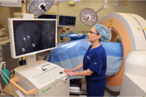
 Dr. Zusman and neurosurgical colleague Dr. Jonathan Forbes made two 1.5-inch incisions on either side of Marge’s spine and operated through small tubes under an operating microscope, using the “O-arm/Intraoperative CT and Stealth Image Guidance” to place pins down the long shaft of the bone, and rods to hold them in place. She was able to clean out the disc space and decompress the pinched nerve roots, placing a spacer, or cage, in the disc space to re-establish its normal height.
“If we had just looked at the MRI images, the abnormality would not have been visible. But we looked at the patient,” said Dr. Zusman.
When Marge took her first steps after surgery, she was stunned. “It was gone. My pain was GONE!” she exclaimed.
Today she can walk a block or more without her legs giving out. She’s thrilled to be able to participate in water therapy at NorthBay HealthSpring Fitness, where therapist Bob Blackwell keeps a close eye on her progress.
“She’ll always be at risk for back health issues, but she knows she needs to stay on top of her health,” said Dr. Zusman, “and she’s committed to doing so.”
Some days, Marge admits she pushes a little too hard. She’s learning where to draw her line in the sand.
But bottom line?
“I am pain-free,” she said emphatically. “I am no longer taking any pain medication for my back. I’m living again and I couldn’t be happier about it. Yes, there was a lot of hard work along the way. I had to follow doctors’ orders and I did. I wore braces, I used my walker, I exercised, I wore a bone stimulator with an electronic pulse. And I listen to my therapists. And thank goodness I did, because it’s all worth it.”
